We use fluorescence microscopy for Histopathological slides imaging and ANA panels. This technique helps detect as few as even 50 molecule per cubic meter.
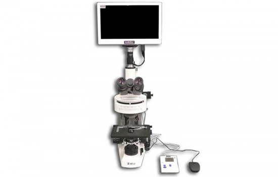
Fluorescence Microscope
- Fluorescence microscopy is the most popular method for studying the dynamic behavior exhibited in live-cell imaging.
- This stems from its ability to isolate individual proteins with a high degree of specificity amidst non-fluorescing material.
- The sensitivity is high enough to detect as few as 50 molecules per cubic micrometer.
- Different molecules can now be stained with different colors, allowing multiple types of the molecule to be tracked simultaneously.
- These factors combine to give fluorescence microscopy a clear advantage over other optical imaging techniques, for both in vitro and in vivo imaging.